 | Info
Sheets |
| | | | | | | | | | | | | | | | | | | | | | | | |
 | Out-
side |
| | | | |
|
| | | | |
Result : Searchterm 'Relaxation Time' found in 5 terms [ ] and 52 definitions [ ] and 52 definitions [ ] ]
| previous 51 - 55 (of 57) nextResult Pages :  [1] [1]  [2 3 4 5 6 7 8 9 10 11 12] [2 3 4 5 6 7 8 9 10 11 12] |  | |  | Searchterm 'Relaxation Time' was also found in the following services: | | | | |
|  |  |
| |
|
( STIR) Also called Short Tau ( t) ( inversion time) Inversion Recovery. STIR is a fat suppression technique with an inversion time t = T1 ln2 where the signal of fat is zero ( T1 is the spin lattice relaxation time of the component that should be suppressed). To distinguish two tissue components with this technique, the T1 values must be different. Fluid Attenuation Inversion Recovery ( FLAIR) is a similar technique to suppress water.
Inversion recovery doubles the distance spins will recover, allowing more time for T1 differences. A 180┬░ preparation pulse inverts the net magnetization to the negative longitudinal magnetization prior to the 90┬░ excitation pulse.
This specialized application of the inversion recovery sequence set the inversion time ( t) of the sequence at 0.69 times the T1 of fat. The T1 of fat at 1.5 Tesla is approximately 250 with a null point of 170 ms while at 0.5 Tesla its 215 with a 148 ms null point. At the moment of excitation, about 120 to 170 ms after the 180┬░ inversion pulse (depending of the magnetic field) the magnetization of the fat signal has just risen to zero from its original, negative, value and no fat signal is available to be flipped into the transverse plane.
When deciding on the optimal T1 time, factors to be considered include not only the main field strength, but also the tissue to be suppressed and the anatomy. In comparison to a conventional spin echo where tissues with a short T1 are bright due to faster recovery, fat signal is reversed or darkened.
Because body fluids have both a long T1 and a long T2, it is evident that STIR offers the possibility of extremely sensitive detection of body fluid. This is of course, only true for stationary fluid such as edema, as the MRI signal of flowing fluids is governed by other factors.
See also Fat Suppression and Inversion Recovery Sequence. | | | |  | | | | | | | | |  Further Reading: Further Reading: | | Basics:
|
|
News & More:
| |
| |
|  | |  |  |  |
| |
|

(SE) The most common pulse sequence used in MR imaging is based of the detection of a spin or Hahn echo. It uses 90┬░ radio frequency pulses to excite the magnetization and one or more 180┬░ pulses to refocus the spins to generate signal echoes named spin echoes (SE).
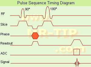
In the pulse sequence timing diagram, the simplest form of a spin echo sequence is illustrated.
The 90┬░ excitation pulse rotates the longitudinal magnetization ( Mz) into the xy-plane and the dephasing of the transverse magnetization (Mxy) starts.
The following application of a 180° refocusing pulse (rotates the magnetization in the x-plane) generates signal echoes. The purpose of the 180° pulse is to rephase the spins, causing them to regain coherence and thereby to recover transverse magnetization, producing a spin echo.
The recovery of the z-magnetization occurs with the T1 relaxation time and typically at a much slower rate than the T2-decay, because in general T1 is greater than T2 for living tissues and is in the range of 100-2000 ms.
The SE pulse sequence was devised in the early days of NMR days by Carr and Purcell and exists now in many forms: the multi echo pulse sequence using single or multislice acquisition, the fast spin echo (FSE/TSE) pulse sequence, echo planar imaging (EPI) pulse sequence and the gradient and spin echo (GRASE) pulse sequence;; all are basically spin echo sequences.
In the simplest form of SE imaging, the pulse sequence has to be repeated as many times as the image has lines. Contrast values:
PD weighted: Short TE (20 ms) and long TR.
T1 weighted: Short TE (10-20 ms) and short TR (300-600 ms)
T2 weighted: Long TE (greater than 60 ms) and long TR (greater than 1600 ms)
With spin echo imaging no T2* occurs, caused by the 180┬░ refocusing pulse. For this reason, spin echo sequences are more robust against e.g., susceptibility artifacts than gradient echo sequences.
See also Pulse Sequence Timing Diagram to find a description of the components.
| | | |  | |
• View the DATABASE results for 'Spin Echo Sequence' (24).
| | | | |  Further Reading: Further Reading: | Basics:
|
|
News & More:
| |
| |
|  | |  |  |  |
| |
|

Image Guidance
Artifacts may appear as a series of fine lines. A narrow bandwidth causes a wide read window, which allows the stimulated echo to be incorporated into the image data. This can be supported by increasing the received bandwidth, which would narrow the read window, thus not incorporating the extraneous echo. Another help would be to change the first echo time, which may change the spacing of the stimulated echoes to outside that of the read window for the second echo. | |  | |
• View the DATABASE results for 'Stimulated Echo' (8).
| | | | |  Further Reading: Further Reading: | Basics:
|
|
| |
|  |  | Searchterm 'Relaxation Time' was also found in the following services: | | | | |
|  |  |
| |
|
( SPIO) Relatively new types of MRI contrast agents are superparamagnetic iron oxide-based colloids (median diameter greater than 50nm). These compounds consist of nonstoichiometric microcrystalline magnetite cores, which are coated with dextrans (in ferumoxide) or siloxanes (in ferumoxsil). After injection they accumulate in the reticuloendothelial system (RES) of the liver (Kupffer cells) and the spleen. At low doses circulating iron decreases the T1 time of blood, at higher doses predominates the T2* effect.
SPIO agents are much more effective in MR relaxation than paramagnetic agents. Since hepatic tumors either do not contain RES
cells or their activity is reduced, the contrast between liver and lesion is improved. Superparamagnetic iron oxides cause noticeable shorter T2 relaxation times with signal loss in the targeted tissue (e.g., liver and spleen) with all standard pulse sequences.
Magnetite, a mixture of FeO and Fe2O3, is one of the used iron oxides. FeO can be replaced by Fe3O4.
Use of these colloids as tissue specific contrast agents is now a well-established area of pharmaceutical development. Feridex┬«, EndoremÔäó, GastroMARK┬«, Lumirem┬«, Sinerem┬«, Resovist┬« and more patents pending tell us that the last word in this area is not said.
Some remarkable points using SPIO:
ÔÇó
A minimum delay of about 10 min. between injection (or infusion) and MR imaging, extends the examination time.
ÔÇó
Cross-section flow void in narrow blood vessels may impede the differentiation from small liver lesions.
ÔÇó
Aortic pulsation artifacts become more pronounced.
See also Superparamagnetism, Superparamagnetic Contrast Agents and Classifications, Characteristics, etc.. | |  | |
• View the DATABASE results for 'Superparamagnetic Iron Oxide' (32).
| | |
• View the NEWS results for 'Superparamagnetic Iron Oxide' (3).
| | | | |  Further Reading: Further Reading: | Basics:
|
|
News & More:
| |
| |
|  | |  |  |  |
| |
|
| |  | |
• View the DATABASE results for 'T1 Relaxation' (18).
| | | | |  Further Reading: Further Reading: | Basics:
|
|
News & More:
| |
| |
|  | |  |  |
|  | | |
|
| |
 | Look
Ups |
| |